具体描述
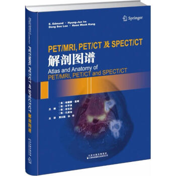
PET/MRI,PET/CT及SPECT/CT解剖图谱
作 者:(美)埃德蒙·基姆(E.Edmund Kim) 等 主编;黄云超 等 译 定 价:280 出 版 社:天津科技翻译出版有限公司 出版日期:2017年08月01日 页 数:478 装 帧:精装 ISBN:9787543336704 ●第1章PET/MRI解剖图谱
●1.1大脑
●1.1.1病例1
●1.1.2病例2
●1.2头颈部
●1.2.1病例1
●1.2.2病例2
●1.3胸部
●1.3.1病例1
●1.3.2病例2
●1.4腹部
●1.4.1病例1
●1.4.2病例2
●1.4.3病例3
●1.4.4病例4
●1.4.5病例5
●1.4.6病例6
●1.4.7病例7
●1.4.8病例8
●1.5盆腔
●部分目录
内容简介
《PET/MRI, PET/CT及SPECT/CT解剖图谱》一书共3章内容,分别为PET/MRI解剖图谱、PET/CT解剖图谱和SPECT/CT解剖图谱。靠前章PET/MRI解剖图谱按照大脑、头颈部、胸部、腹部、盆腔、肌肉骨骼系统进行描述。第2章PET/CT解剖图谱按照FDG和非-FDG进行分类描述。第3章则描述了肿瘤、骨和其他病变的SPECT/CT表现。该书内容全面、新颖,配有大量高清图片,实用性较强,可供核医学科、放射科、肿瘤科、神经科及心内科等相关医师参考阅读。 (美)埃德蒙·基姆(E.Edmund Kim) 等 主编;黄云超 等 译 埃德蒙·基姆,加利福尼亚大学欧文分校放射系教授,首尔国立大学和庆熙大学分子医学系教授。
任亨浚,首尔国立大学医院核医学系讲师。
李东洙,首尔国立大学医院核医学系教授。
孔建旭,首尔国立大学医院核医学系主任、教授。
黄云超,男,教授,昆明医学院第三附属医院、昆明医学院临床肿瘤学院、云南省肿瘤医院副院长、云南省肿瘤生物免疫治疗中心副主任、肿瘤研究所副所长。担任中华医学会云南省胸心血管外科分会副主任委员,中国抗癌协会肺癌专业委员会常委,云南省抗癌协会肺癌专业委员会主任委员。
孙华,主任医师,云南省肿瘤医院暨昆明医科大学第三附属医院PET/CT中心主任,九三学社等
PET/MRI, PET/CT and SPECT/CT Anatomy Atlas: A Comprehensive Guide for Imaging Interpretation Introduction In the rapidly evolving landscape of medical imaging, the integration of functional and anatomical information has revolutionized diagnostic capabilities. Positron Emission Tomography (PET), Computed Tomography (CT), Magnetic Resonance Imaging (MRI), and Single-Photon Emission Computed Tomography (SPECT) are powerful modalities that, when combined, offer unprecedented insights into disease processes. This atlas, "PET/MRI, PET/CT and SPECT/CT Anatomy Atlas," is meticulously designed to serve as an indispensable resource for clinicians and researchers seeking to master the interpretation of these advanced hybrid imaging techniques. It bridges the gap between the intricate anatomy visualized by CT and MRI and the metabolic and functional data provided by PET and SPECT, enabling a more precise and comprehensive understanding of normal anatomy and pathological alterations. Scope and Objectives This atlas focuses on the anatomical representation within the context of PET/MRI, PET/CT, and SPECT/CT imaging. Its primary objective is to equip readers with a profound understanding of how anatomical structures appear on these combined modalities, allowing for accurate localization and characterization of functional abnormalities. We delve into the characteristic attenuation and signal properties of various tissues and organs as depicted by CT and MRI, and how these correlate with the distribution of radiotracers in PET and SPECT. The atlas aims to: Provide detailed anatomical atlases: Presenting a systematic, region-by-region approach to the human body, covering all major organ systems. Illustrate normal anatomical variations: Highlighting common variations in anatomy that can be mistaken for pathology, thereby enhancing diagnostic accuracy. Explain the principles of hybrid imaging: Briefly touching upon the underlying physics and technical considerations that contribute to the fused image appearance. Correlate anatomical structures with functional information: Demonstrating how PET and SPECT tracers highlight specific physiological processes within well-defined anatomical boundaries. Offer practical guidance for interpretation: Presenting examples and common pitfalls to aid in the daily interpretation of PET/MRI, PET/CT, and SPECT/CT scans. Target Audience This atlas is tailored for a wide range of medical professionals involved in the acquisition, interpretation, and application of advanced hybrid imaging techniques. This includes, but is not limited to: Radiologists: Seeking to enhance their understanding of fused imaging interpretation and its anatomical correlates. Nuclear Medicine Physicians: Requiring detailed anatomical knowledge to precisely localize and interpret functional findings. Oncologists: Utilizing these modalities for cancer staging, treatment response assessment, and recurrence detection. Neurologists and Neurosurgeons: Employing PET/MRI and PET/CT for the evaluation of neurological disorders, including tumors, epilepsy, and degenerative diseases. Cardiologists: Using these techniques for the assessment of myocardial perfusion, viability, and inflammatory conditions. Researchers: Investigating novel radiotracers and their anatomical distribution in various physiological and pathological states. Trainees and Residents: Building a foundational knowledge of hybrid imaging anatomy. Content Structure and Approach The atlas is organized in a logical and systematic manner, progressing from general principles to specific anatomical regions. Each chapter is dedicated to a particular area of the body, ensuring a comprehensive and thorough exploration. Within each section, we adopt a multi-faceted approach: 1. Introduction to the Region: Briefly outlining the anatomical boundaries and key structures of the region. 2. CT/MRI Anatomy Review: Providing a detailed depiction of the anatomical structures as seen on conventional CT and MRI, emphasizing their typical appearance, signal intensity, and attenuation characteristics. This section serves as the foundational anatomical reference. 3. PET/SPECT Tracer Distribution in Normal Anatomy: Illustrating how common PET and SPECT radiotracers distribute in normal physiological states within the discussed anatomical structures. This section highlights the metabolic landscape of healthy tissues. We will discuss tracers such as [¹⁸F]FDG, [¹¹C]choline, [¹⁸F]PSMA, [¹²³I]ioflupane, [⁹⁹mTc]MDP, and others relevant to each region. 4. PET/CT, PET/MRI, and SPECT/CT Fused Image Representation: This is the core of the atlas. We present meticulously selected fused images, showcasing the co-registration of functional data (PET/SPECT) with anatomical information (CT/MRI). Emphasis will be placed on identifying specific anatomical landmarks and correlating them with the intensity and distribution patterns of the radiotracers. This section will demonstrate how to discern normal anatomical structures that might otherwise appear as "hot" or "cold" spots on functional imaging alone. 5. Common Anatomical Variations and Pitfalls: Addressing frequently encountered anatomical variations that can mimic pathology on hybrid imaging, providing guidance on how to differentiate them from true abnormalities. This section is crucial for avoiding misinterpretations. 6. Pathological Examples (Briefly Referenced): While not a pathology atlas, brief references to how specific pathologies manifest within the context of anatomical localization will be made, reinforcing the importance of accurate anatomical identification for diagnostic purposes. Detailed Content Outline (Sample Chapters) Chapter 1: Head and Neck Introduction: Boundaries of the head and neck, key structures (brain, orbits, salivary glands, thyroid, larynx, pharynx, major vessels). CT/MRI Anatomy: Detailed views of cranial bones, brain parenchyma (gray matter, white matter, cerebrospinal fluid), cranial nerves, vascular structures, orbits, nasal cavity, paranasal sinuses, oral cavity, pharynx, larynx, thyroid gland, parathyroid glands, and cervical lymph nodes. Emphasis on contrast enhancement patterns and signal characteristics. PET/SPECT Tracer Distribution (Normal): Brain: Glucose metabolism ([¹⁸F]FDG), neurotransmitter systems ([¹¹C]raclopride, [¹⁸F]flutemetamol for amyloid, [¹⁸F]THK-5351 for tau). Salivary Glands: Functional assessment of secretory activity. Thyroid: Iodine uptake ([¹²³I]sodium iodide). Head and Neck Tumors: Metabolic activity ([¹⁸F]FDG, [¹¹C]choline, [¹⁸F]PSMA). Fused Image Representation: Detailed atlases of axial, coronal, and sagittal PET/CT, PET/MRI, and SPECT/CT slices of the head and neck, highlighting the precise anatomical localization of radiotracer uptake. Examples of basal ganglia, cortex, cerebellum, cranial nerves, major arteries and veins, thyroid gland, and salivary glands. Anatomical Variations/Pitfalls: Variations in venous drainage, anatomical variants of cranial nerves, normal parathyroid gland visibility, residual thyroid tissue after treatment. Chapter 2: Thorax Introduction: Boundaries of the thorax, key structures (lungs, pleura, mediastinum, heart, great vessels, esophagus). CT/MRI Anatomy: Detailed views of lung lobes and segments, airways (trachea, bronchi), pleura, mediastinal compartments, heart chambers and valves, pericardium, aorta, pulmonary arteries and veins, superior vena cava, inferior vena cava, esophagus, and thoracic spine. PET/SPECT Tracer Distribution (Normal): Lungs: Alveolar gas exchange, lung perfusion. Heart: Myocardial perfusion ([⁹⁹mTc]sestamibi, [¹⁸F]FDG for viability), cardiac sympathetic innervation. Mediastinum: Lymph node uptake patterns. Fused Image Representation: Comprehensive atlases of axial, coronal, and sagittal PET/CT, PET/MRI, and SPECT/CT slices of the thorax. Emphasis on visualizing the lungs, mediastinal structures (thymus, lymph nodes, great vessels), heart, and thoracic spine. Correlation of [¹⁸F]FDG uptake in respiratory muscles and airways. Anatomical Variations/Pitfalls: Variations in bronchial anatomy, common aortic arch branching patterns, normal thymus appearance in children and adults, diaphragmatic position, costophrenic angle variations. Chapter 3: Abdomen Introduction: Boundaries of the abdomen, key organs (liver, spleen, pancreas, kidneys, adrenal glands, gastrointestinal tract, major vessels). CT/MRI Anatomy: Detailed depiction of the liver lobes and segments, gallbladder and biliary tree, spleen, pancreas (head, body, tail), kidneys (cortex, medulla, pelves), adrenal glands, stomach, small intestine, large intestine, aorta, inferior vena cava, renal arteries and veins, and retroperitoneal lymph nodes. PET/SPECT Tracer Distribution (Normal): Liver: Hepatobiliary clearance, metabolic activity. Spleen: Functional assessment. Kidneys: Glomerular filtration rate, tubular function. Gastrointestinal Tract: Baseline metabolic activity, specific tracer uptake in certain conditions. Fused Image Representation: Extensive atlases of axial, coronal, and sagittal PET/CT, PET/MRI, and SPECT/CT slices of the abdomen. Focusing on precise anatomical localization of the liver, spleen, pancreas, kidneys, adrenal glands, and segments of the gastrointestinal tract. Demonstrating normal physiological radiotracer uptake in these organs. Anatomical Variations/Pitfalls: Variations in liver lobar segmentation, accessory spleens, common variations in pancreatic duct anatomy, renal vascular variations, congenital anomalies of the gastrointestinal tract, retroperitoneal lymph node distribution. Chapter 4: Pelvis Introduction: Boundaries of the pelvis, key organs (bladder, rectum, prostate, seminal vesicles, uterus, ovaries, pelvic floor, major vessels, lymph nodes). CT/MRI Anatomy: Detailed visualization of the bladder, rectum, prostate gland (zones), seminal vesicles, uterus, cervix, vagina, ovaries, fallopian tubes, pelvic bones, pelvic musculature, and pelvic lymph node chains. PET/SPECT Tracer Distribution (Normal): Prostate: Specific tracer uptake ([¹⁸F]PSMA, [¹¹C]choline). Bladder: Normal physiological excretion. Pelvic Lymph Nodes: Baseline metabolic activity. Fused Image Representation: Comprehensive atlases of axial, coronal, and sagittal PET/CT, PET/MRI, and SPECT/CT slices of the pelvis. Precise anatomical delineation of the pelvic organs and their relationship to surrounding structures. Illustrating normal radiotracer distribution, particularly focusing on prostate imaging with specific tracers. Anatomical Variations/Pitfalls: Variations in prostate gland size and shape, anatomical variations of the seminal vesicles, uterine anomalies, pelvic lymph node station variations. Chapter 5: Musculoskeletal System Introduction: Bones, joints, muscles, and associated soft tissues. CT/MRI Anatomy: Detailed imaging of major bones, joints, ligaments, tendons, and musculature of the extremities and axial skeleton. Emphasis on cross-sectional anatomy. PET/SPECT Tracer Distribution (Normal): Bone Scintigraphy ([⁹⁹mTc]MDP): Normal bone turnover, epiphyseal plates in children. Soft Tissue Imaging: Metabolic activity in muscles. Fused Image Representation: Atlases of PET/CT and SPECT/CT images of limbs and axial skeleton, focusing on the anatomical localization of bone and soft tissue. Demonstrating how to interpret physiological bone tracer uptake and distinguish it from pathology. Anatomical Variations/Pitfalls: Variations in ossification centers, muscle origins and insertions, common joint capsule variations. Key Features and Benefits High-Quality Imaging: Utilizes excellent quality, representative fused images from state-of-the-art PET/CT, PET/MRI, and SPECT/CT scanners. Clear Anatomical Labeling: All anatomical structures are meticulously labeled for easy identification. Correlative Imaging: Seamless integration of anatomical and functional information, demonstrating how to interpret the fused images effectively. Focus on Normal Anatomy: Prioritizes the understanding of normal anatomical variations and physiological tracer distribution to provide a strong foundation for pathological interpretation. Practical Approach: Offers actionable insights and guidance for the daily interpretation of hybrid imaging studies. Comprehensive Coverage: Encompasses the entire human body, from the head to the pelvis and musculoskeletal system. Conclusion "PET/MRI, PET/CT and SPECT/CT Anatomy Atlas" is an invaluable educational tool designed to empower clinicians with the knowledge and skills necessary to confidently interpret complex hybrid imaging data. By providing a detailed and systematic exploration of normal anatomy as visualized on these advanced modalities, this atlas will significantly enhance diagnostic accuracy, improve patient management, and contribute to the advancement of medical imaging interpretation. It stands as a testament to the crucial role of anatomical understanding in unlocking the full potential of functional imaging.


